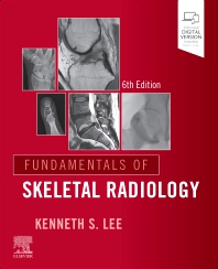
Description
Kenneth S. Lee’s sixth edition introduces the concepts used to understand and interpret skeletal imaging. Radiographs, MRI, CT, ultrasound, and bone-scan examples illustrate musculoskeletal findings, with particular attention to MRI of the spine and joints. Concise explanations connect imaging methods with common pathology and interpretation.
Topics covered
- Core principles for reading skeletal and musculoskeletal images.
- Recognising patterns and narrowing diagnostic possibilities.
- Illustrated explanations supporting an introductory approach to the subject.
Who it’s for
Radiology residents, medical students, and clinicians starting or refreshing their study of skeletal radiology.
Study use: Read a topic’s explanation before comparing its images, then practise describing the features that support the diagnostic pattern.
Reference details
- Authors / editors
- Kenneth S. Lee
- Publisher
- Elsevier
- Edition / publication
- 6th edition
Publisher reference for this title
Choosing this resource
A foundational skeletal imaging text suited to building an approach before consulting a more extensive disease-specific reference.


