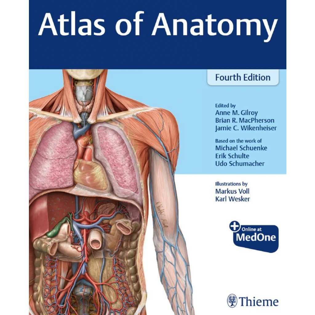
Description
The fourth edition presents human anatomy through a large collection of labelled illustrations and supporting tables. Expanded radiological material connects anatomical structures with radiographs, CT, and MRI, while clinical boxes relate anatomy to function, disease, and variation. Its head and neck organisation reflects a dissection-based approach to learning.
Topics covered
- Regional anatomy presented through detailed labelled illustrations.
- Spatial relationships between muscles, vessels, nerves, and organs.
- Clinical correlations and radiological images linking anatomy with practice.
Who it’s for
Medical and health science students, anatomy learners, and clinicians refreshing regional anatomy.
Study use: Identify structures on the atlas illustrations, then test your understanding of their relationships during dissection or image-based review.